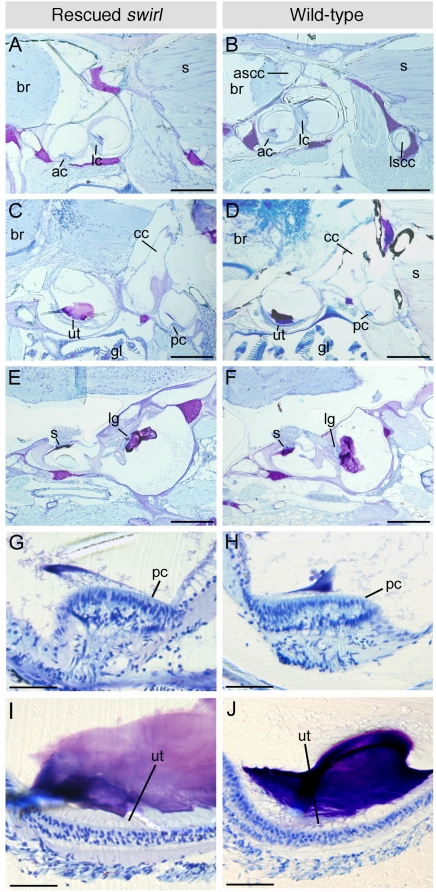
Figure 1. Semicircular canal ducts are absent, but sensory patches are present, in the inner ears of adult rescued swr zebrafish.
(A–F) 10 µm resin parasagittal sections through the inner ears of adult rescued swr zebrafish and age-matched wild-types (anterior to the left, dorsal to the top). Note the absence of semicircular canal ducts in the rescued swr ear (A), which are clearly present in the wild-type ear in an equivalent section (B, showing lumens of anterior and posterior canal ducts). All sensory patches detected in the wild-type ears are also present in the rescued swr ears. Differences in the orientation of the lagenar macula (E, F) reflect a slight difference in the position of the wild-type and swr sections for this pair of panels. (G–J) Higher magnification views of the posterior crista (G, H) and utricular macula (I, J). The structure of the sensory patches is similar in both rescued swr and wild-type ears. Abbreviations: ac, anterior crista; lc, lateral crista; pc, posterior crista; ascc, anterior semicircular canal duct; lscc, lateral semicircular canal duct; cc, crus commune; ut, utricular macula; s, saccular macula; lg, lagenar macula; br, brain; s, somite; gl, gill. Scale bar, (A–F) 400 µm; (G–J) 50 µm.

