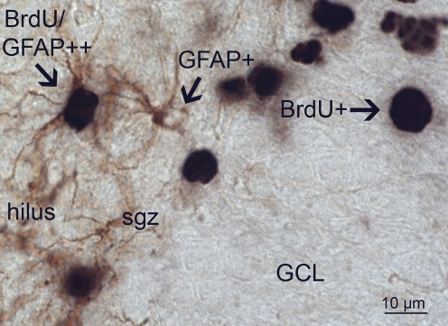
Figure 9. GFAP/BrdU double labeling.
Immunohistochemical double labeling for GFAP and BrdU shows single GFAP+ astrocytes in the hilus with their processes occasionally extending into the sgz. GFAP+ cells reveal brown DAB-staining in their processes and cytoplasm whereas the nucleus is devoid of staining. In the GCL, BrdU+ single cells are stained black by DAB-nickel as indicated (BrdU+) in the granular cell layer (GCL). Shown on the left is a BrdU/GFAP double labeled cell (arrow).

