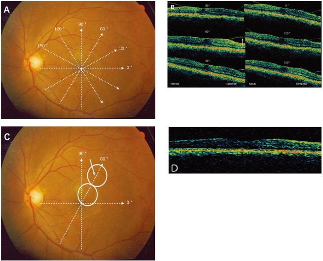
Fig. 1.
Localization of the retinal opening site with OCT retinal mapping. (A) Preoperative OCT retinal mapping of patient 4 (50-year-old female, left eye) contains radial spoke pattern of six scans 6 mm long, centered on the patient's fixation point. (B) Cross-sectional optical coherence tomograms obtained from the corresponding six radial scans. A blue arrow points to the site with posterior hyaloid detachment farthest from the retina. (C) During the operation, two imaginary circles the size of the optic nerve head are drawn to approximate a length of 3 mm. The blue arrow within the second circle points to the opening site for an access to subhyaloid space. (D) Three months after the surgery, the foveal detachment has resolved and the foveal pit is restored. The OCT is taken at 60 degree plane.

