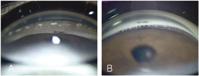
Fig. 2.
Width of iridocorneal angle at 9 months after ICL surgery. (A) Peripheral iris was very steep and convex. Iridocorneal angle was narrowed because of the obstruction of LI sites. (B) After additional LI, although the angle width increased, trabecular pigment also increased. 25 ×, Goldmann 3 mirrors.

