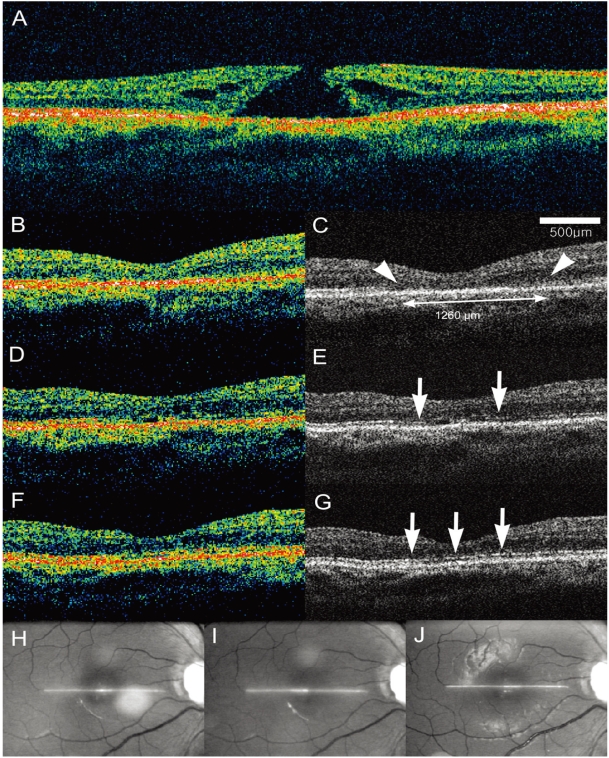
Fig. 3.
Case 3. Serial horizontal optical coherence tomography (OCT) scans after successful macular hole surgery. (A) Preoperative OCT scan shows a stage 2 macular hole. (B and C) Conventional pseudocolor image and enhanced image at 2 weeks demonstrate disorganization of the outer retina (arrow heads). The width was measured as 1260 µm. (D and E) At 6 months, OCT images disclose the partly reorganized photoreceptor layer, which appeared as a broken line on the enhanced image (arrows). (F and G) At 1 year, further reorganization of the photoreceptor layer is identified (arrows). Visual acuity improved gradually from 20/100 (2 weeks) to 20/80 (6 months) and then to 20/40 (1 year). (H, I, and J) The identical location of the OCT scans was verified based on the video images taken simultaneous.

