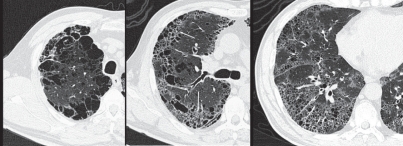
Figure 27.
Combined pulmonary fibrosis and pulmonary emphysema (CPFE) in a 59-year-old male who was a heavy smoker: High resolution computed tomography images.
In the upper lung field (a), a prominent bullous change is apparent. In the middle lung field (b), tiny air cysts (arrows) with ground-glass opacity are present in addition to the bullous changes. In the lower lung field (c), distributed tiny air cysts with definable walls and ground-glass opacity are apparent. These features are consistent with interstitial fibrosis rather than pulmonary emphysema.

