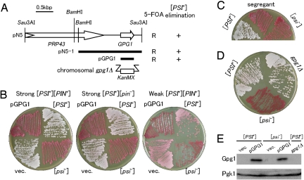
Fig. 1.
Over-expression of GPG1 eliminates the [PSI+] prion. (A) Schematic representation of genes cloned in plasmid pN5 and its derivatives. Open arrows identify protein-coding sequences. The bold bars indicate segments subcloned in constructing plasmids pN5–1 and pGPG1. The ability of these clones to confer 5-FOA resistance (R) and red color (i.e., [psi−]) (+) on a [PSI+] strain (NPK265; ade1–14 ura3–197) is indicated on the right. The gpg1Δ denotes a substitution of the KanMX marker for the chromosomal GPG1 gene. (B) [PSI+] elimination by the Gpg1-expressing plasmid, pGPG1. Strong or weak [PSI+] cells (ade1–14) were transformed with an empty vector (vec.: pRS413, single-copy plasmid with HIS3 marker) or pGPG1. Transformants were selected on SC-his after 3 days and regrown on YPD for 4 days to monitor colony color. [PSI+] and [psi−] control cells are also shown. Strains: Left, NPK265 (strong [PSI+] [PIN+]); Middle, NPK50 (strong [PSI+] [pin−]); Right, NPK197 (weak [PSI+] [pin−]). (C) Complete curing of [PSI+] after pGPG1 plasmid segregation. [PSI+] (NPK265 [PIN+]) cells were transformed with pGPG1 as shown in Fig. 1B (Left), and isolates that had spontaneously lost the plasmid were grown on YPD for 4 days. The “red” colonies remained red even in the absence of the pGPG1 plasmid. (D) Effect of chromosomal gpg1Δ on [PSI+] phenotype. Chromosomal GPG1 allele of NPK265 was nullified by substitution with the KanMX marker (strain BY4742 gpg1Δ) and colony color was monitored. (E) Gpg1 is visualized by immunoblotting. Equal amounts of total protein from whole cell lysates of NPK265 ([PSI+]) and its [psi−] derivative carrying plasmid pGPG1 or an empty vector (vec.), as well as from NPK563 (gpg1Δ), were separated by SDS/PAGE and immunoblotted with anti-Gpg1 antibody.

