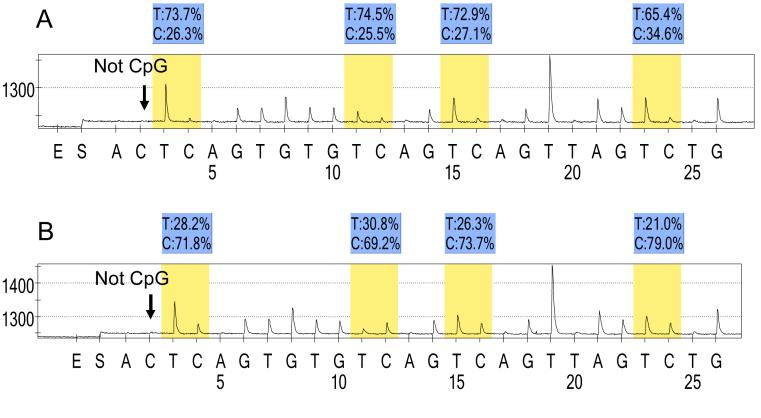
Figure 1. Pyrosequencing for LINE-1 methylation.
A. MSS CIMP-low tumor. B. MSI-high CIMP-high tumor. The % numbers (in blue shade) are proportions of C and T at each CpG site after bisulfite conversion. Thus, the methylation level of each CpG site is estimated by the proportion of C (%). An overall LINE-1 methylation level is calculated as the average of the proportions of C (%) at the 4 CpG sites. The first, third and fourth CpG sites follow stretches of Ts, resulting in higher T peaks (in yellow shade) than the second CpG site, and the proportion of C (%) has been adjusted accordingly. The arrows indicate no residual C at the non-CpG site, ensuring complete bisulfite conversion.
CIMP, CpG island methylator phenotype; MSI, microsatellite instability; MSS, microsatellite stable.

