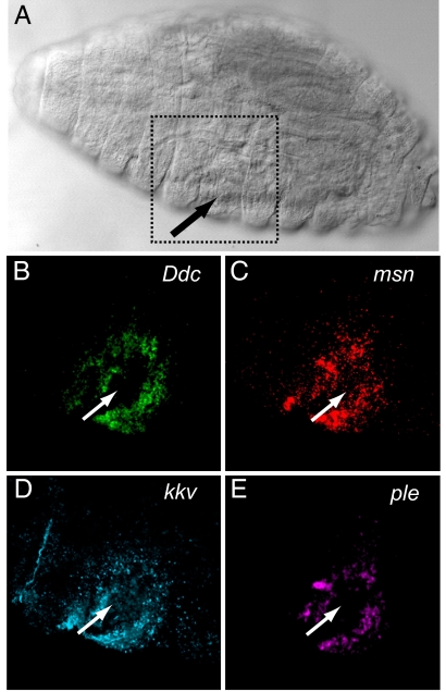
Fig. 4.
Ddc, ple, msn, and kkv are transcriptionally activated in epidermal cells around wounds. (A) Image of wounded embryo, visualized with DIC optics, fixed 30 min after wounding. Arrow shows entry wound; dotted box shows the region imaged with fluorescence optics in frames B–E. (B–E) Ddc (B), msn (C), kkv (D), and ple (E) transcripts were simultaneously detected in the embryo around the aseptic wound using hapten-labeled probes (35). No signals were detected around wounds in embryos fixed immediately after wounding and hybridized with probes (data not shown).

