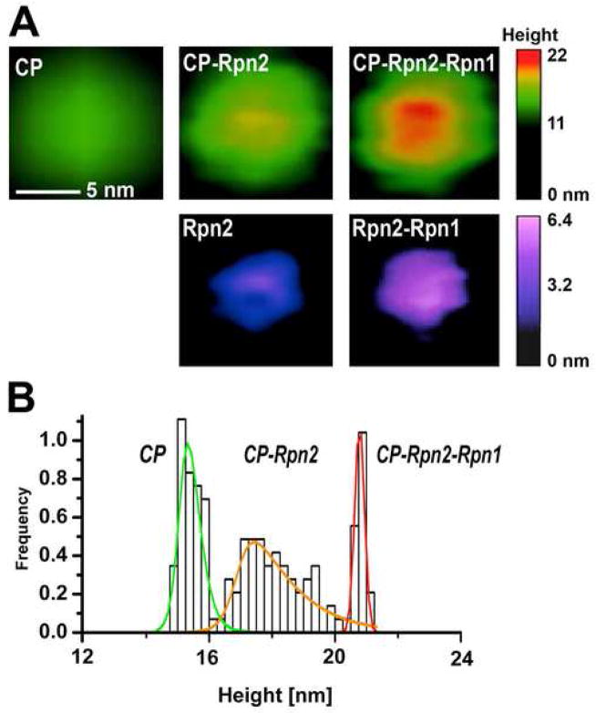
Fig. 1. “Building up” the proteasome assembly.
The particles electrostatically adsorbed to mica were imaged in tapping mode in liquid (Nanoscope IIIa; Veeco). (A) Representative top-view images of 20S core particle (CP) and core particle extended with a “stent” created from Rpn2 or Rpn1-Rpn2 dimer, presented below. The images were zoomed-in from 600 nm × 600 nm fields, flattened and plain-fitted. Color bars represent the height scale, different for CP and free stent components. (B) Height distribution of 20S CP in mixture with Rpn1 and Rpn2 is shown as relative number of molecules, which fall within a height range of 0.25 nm wide bins. Heights were analyzed with grain analysis function (SPIP v. 4.3.2.0, Image Metrology, Denmark). To fit the experimental frequency data, normal distribution of particle height was assumed for each type of particles, with the exception of the broad peak (presumably mostly CP-Rpn2), for which the height distribution curve was fit using an asymmetric double sigmoidal function within the OriginPro Peak Fitting module (modified from Rosenzweig, Osmulski, Gaczynska, Glickman, submitted).

