
Fig. 5. Detection of the surface (A) and cytosolic (B) Annexin II expression in IEC-18 cells by flow cytometry.
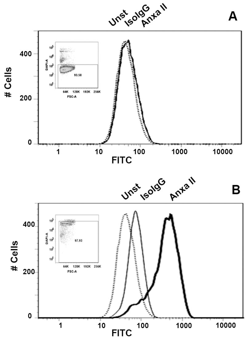
Cells were detached with 0.5 mM EDTA, and the cell suspension was directly used for labeling (A), or initially fixed and permeabilized by PFA (1.6%) and cold methanol, respectively, (B). Aliquots (106) of the cell suspensions were left untreated (Unst; dotted line) or incubated with the fluorescein-conjugated anti-Annexin II (ANXA II; heavy line) or isotype-matched (IsoIgG; fine line) antibodies. Cells were washed and resuspended in DAPI-containing buffer prior to analysis. Inserts show DAPI fluorescence (Y- axis) versus forward light scattering characteristics of the analyzed populations.