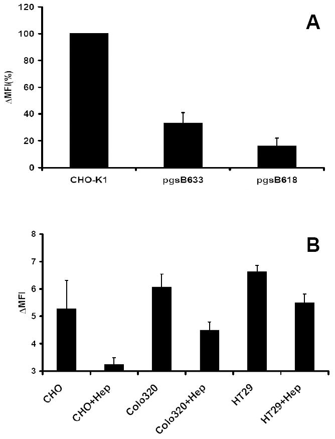
Fig. 7. Bio-PG binding is dependent on surface proteoglycans.

(A). The wild type and proteoglycan-deficient strains of CHO-K1 cells were analyzed for bio-PG (100 nM) binding by flow cytometry as above, except that the binding reaction was performed in HBSS- 1% BSA. The Mean Fluorescence Intensity was calculated and presented as % of MFI for wild type CHO-K1 cells (MFI=100%). The figure shows combined data from three separate experiments. (B). Heparinase pre-treatment attenuates bio-PG to epithelial cells. The indicated cells were treated (+Hep) or not with heparinase (1000U/ml) for 1.5 hours at 37°C before testing bio-PG binding as above. Data (in triplicate) for one of two performed experiments. Statistically significant (P<0.05) decrease of bio-PG binding was found in treated (+Hep) versus untreated samples.
