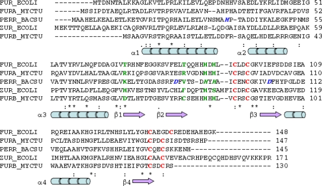
Figure 2.
Sequence alignment of Furec with FurA, PerRbs together with Zurec and FurB (performed with ClustalW2 [45]). Residues of the putative regulatory binding site identified in the crystal structure of FurB are depicted in green, residues of the putative structural binding site are red [43]. The proposed binding residues for the second, regulator binding site in PerRbs His37, Asp85, His91, His 93 and Asp104 are shown in blue [46]. Secondary structure assignment is based on the FurB crystal structure: blue rods refers to α-helix, and violet arrows to β-strands. The last line shows sequence conservation: '*' denote conserved residues, '.' and ':' indicate similar residues in the alignment.

