
Figure 5. The C1 domain of AC6 most strongly localizes in lipid raft/caveolar fractions.
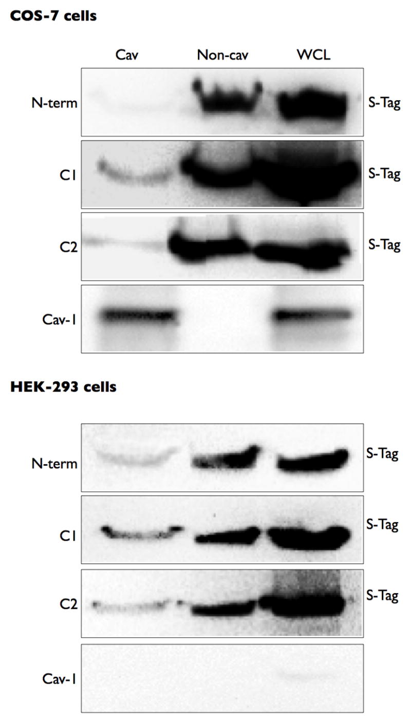
COS-7 or HEK-293 cells were transfected with each intracellular domain construct. Cells were then fractionated using a non-detergent method to isolate lipid rafts and caveolae (see Methods). Following sucrose density centrifugation, fractions were 2–4 were combined as the Cav fraction and fractions 7–10 were combined as the non-cav fraction. Each pooled fraction and a whole cell lysate control were separated by SDS-PAGE and analyzed by immunoblot using an anti-His antibody to detect the expressed protein construct. Images shown are representative of 4–5 experiments.