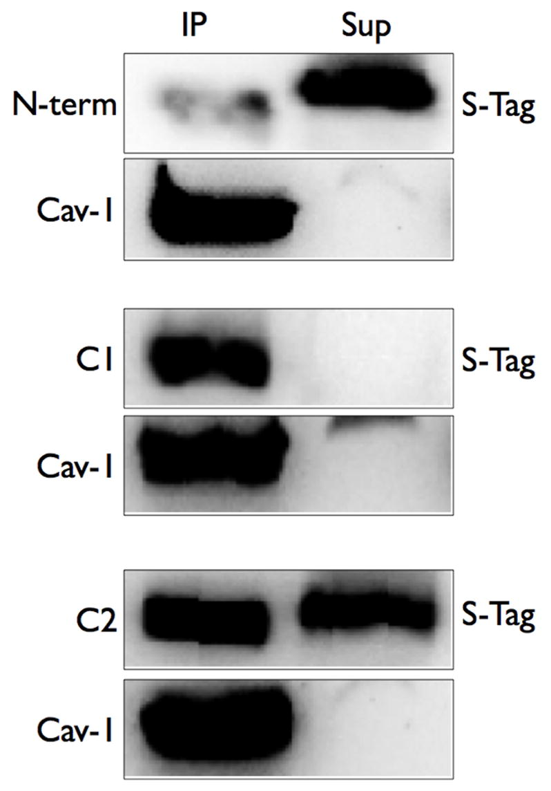
Figure 6. The C1 and C2 domains are localized in Triton X-100 insoluble fractions and immunoprecipitate with caveolin-1.

COS-7 cells were transfected with each intracellular domain construct. Cells were then separated into soluble and insoluble fractions in Triton-X 100 (left panels) or were lysed and immunoprecipitation of caveolin-1 was performed (right panels, see Methods). Soluble and insoluble fractions or caveolin-1 immunoprecipitates (IP) or supernatants (Sup) were separated by SDS-PAGE and analyzed by S-Tag detection (to detect AC6 domain proteins) or immunoblotting (to detect caveolin-1). Images shown are representative of 5 experiments.
