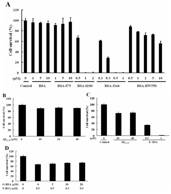
Figure 3.
Cytotoxic effect of Fibrillar BSAs. Cell viability was determined by the MTT assay. (A) BHK-21 cells were treated for 8 h with various concentrations of G-BSA, BSA-S75, BSA-S200, BSA-Zwit, or BSA-HW55S as indicated in serum-free medium. (B) BHK-21 cells were treated with increasing concentrations of Aβ25–35 in serum-free medium for 8 h. (C) BHK-21 cells were treated with increasing concentrations of Aβ25–35 or F-BSA (BSA-S200) in serum-free medium for 24 h. (D) BHK-21 cells were treated with or without increasing concentrations of G-BSA (BSA) for 1 h, then incubated with 0.5 μM F-BSA (BSA-S200) in serum-free medium for 8 h. Data are means ± S.D. (n = 3) of percentage of cell survival as determined by the MTT assays.

