
Figure 6.
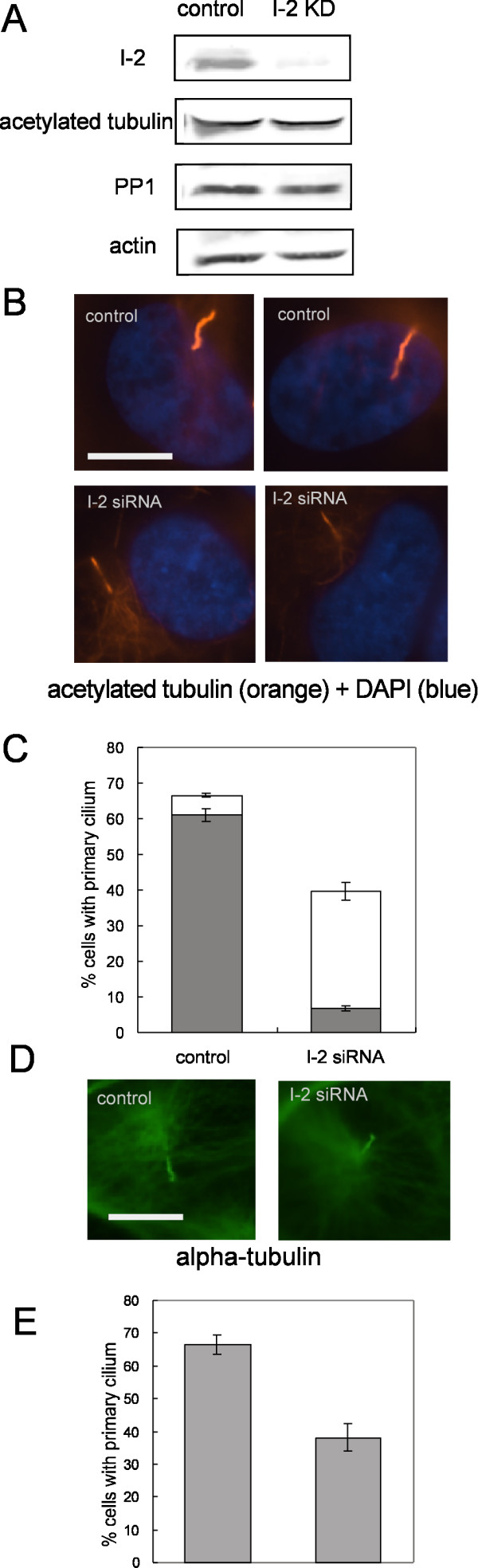
Knockdown of I-2 reduces axonemal alpha-tubulin acetylation and primary cilium assembly. (A) Cells were incubated with control siRNA or I-2 siRNA at 24 hrs before confluence, and collected 48 hrs after confluence. Extracts were subjected to immunoblotting as described in Methods. Actin was used as loading control. (B) ARPE-19 cells transfected with control siRNA or I-2 siRNA were fixed and stained for acetylated-tubulin (orange) and DNA (blue). Scale bar = 10 micron. (C) Quantitation of cells with a primary cilium. Cells with a primary cilium evidenced by acetylated-tubulin staining were scored in 200 cells per group. Intensity of tubulin acetylation in the primary cilium was measured as described in Methods. Solid bars represent percentage of cells possessing a primary cilium with full level of alpha-tubulin acetylation, whereas open bars represent percentage of cells possessing a primary cilium with reduced levels of tubulin acetylation. Data were plotted as mean ± SD of values from three independent experiments. (D) Cells transfected with control siRNA or I-2 siRNA were fixed and stained for alpha-tubulin (green). Scale bar = 10 micron. (E) Percentage of cells with a primary cilium based on alpha-tubulin staining was scored in 200 cells per group. Results were plotted as mean ± SD of values from three independent experiments.