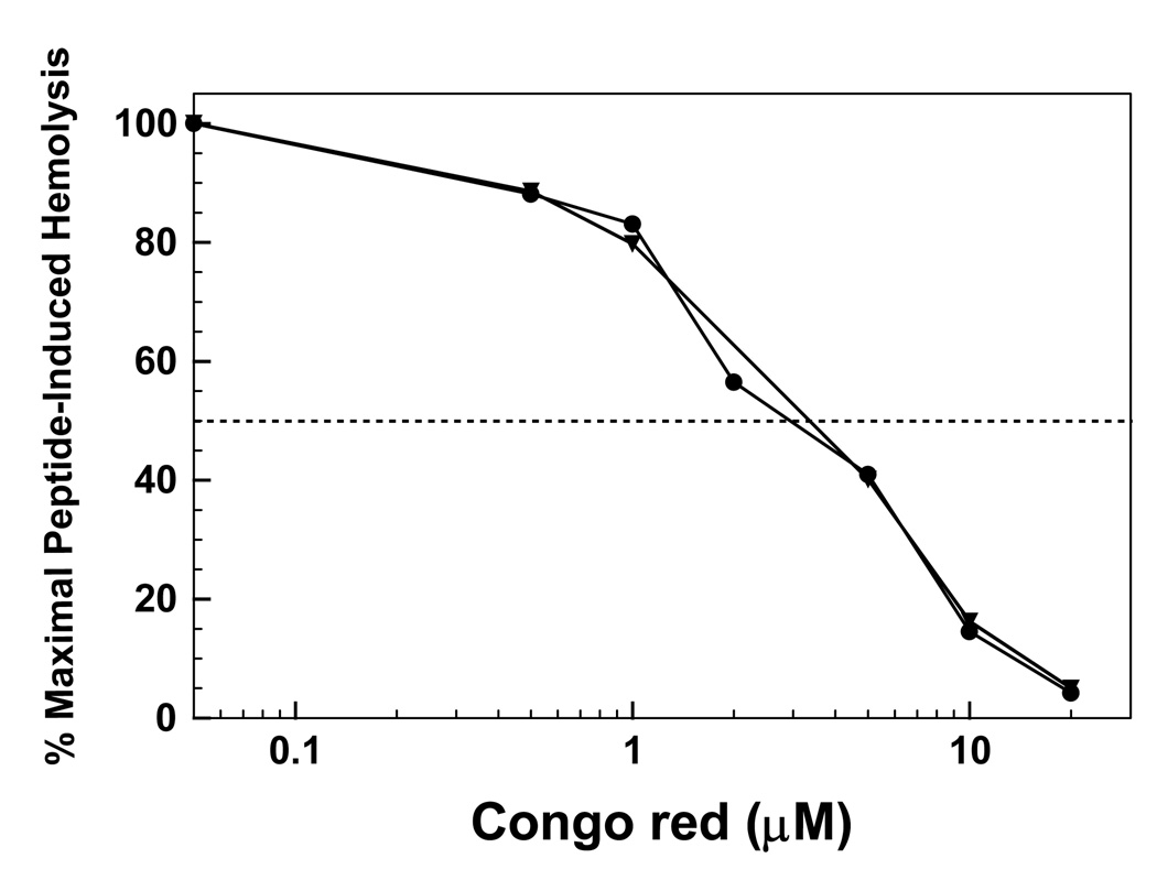
fig. 10.
Congo red (CR) inhibition of hemolysis induced by FP (●) or Aβ(26–42) (▼). Erythrocytes (1 × 10¹¹ cells/liter) were incubated with 60 µM FP or Aβ(26–42), and the indicated concentrations CR for 30 min at 37°C. Percent hemolysis was determined as described in Section 2, and is reported as the mean of duplicate determinations. One hundred percent maximal hemolysis for the inhibition curves is defined as that obtained by incubating 60 µM FP or 60 µM Aβ(26–42) with 10 µl of packed erythrocytes in 0.5 ml of PBS, without inhibitor. The dashed horizontal line represents 50% of the maximal hemolysis, observed without CR.

