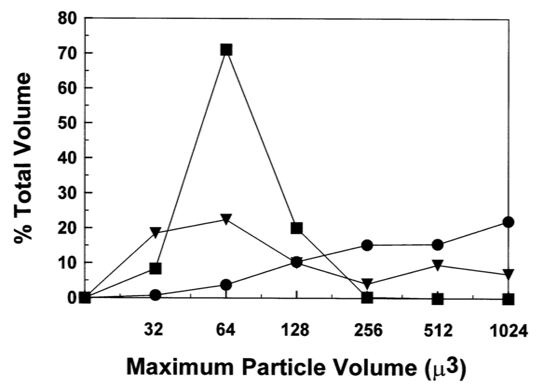
fig. 7.
Coulter counter sizing of erythrocytes treated with HIV-1 FP and Aβ(26–42). Erythrocytes were tested for aggregation by incubating 10 µl of packed red blood cells, diluted to 0.5 ml in PS buffer, with the following agents at 37°C for 30 min: 60 µM FP (●); 60 µM Aβ(26–42) (▼); control PBS (■). At the end of each incubation, an aliquot of the reaction mixture was diluted 1/500 in isotonic phosphate-buffered saline. The number of particles in each size range (i.e., maximum particle volume µ3,x axis) shown was then measured with a Coulter counter. Percent (total volume) for the particles in each channel (y-axis) is calculated as in Section 2. Results are reported as the mean of duplicate determinations, and are representative of three independent experiments.

