Figure 3.
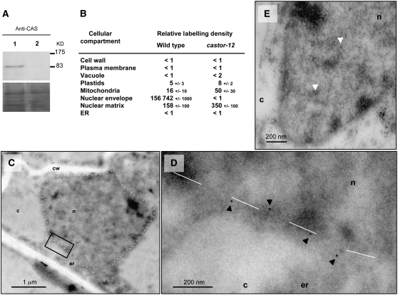
Immunogold Localization of CASTOR in L. japonicus with Anti-CASTOR.
(A) Immunoblot detection with anti-CASTOR in total extract of transformed roots of L. japonicus mutant castor-12 (W93*). Lane 1, castor-12 complemented with P35S:CASTOR via A. rhizogenes hairy root transformation; lane 2, castor-12 transformed roots with empty vector as a negative control. Bottom panel, loading shown by Coomassie blue staining.
(B) Relative density of gold particles in different cellular compartments of L. japonicus Gifu wild-type and mutant castor-12 roots after immunogold labeling with anti-CASTOR.
(C) to (E) Electron microscopic images of L. japonicus Gifu wild-type nucleus (C) and castor-12 nucleus (E). (D) shows an enlargement of the black box in (C). Dashed white line, delimitation of the nuclear envelopes. c, cytoplasm; cw, cell wall; er, endoplasmic reticulum; n, nucleus. Black arrowheads mark positive immunodetection of CASTOR, and white arrowheads mark background labeling in the nuclear matrix.

