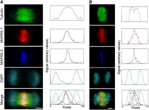
Figure 3.
Comparison of MAP65-1 and MAP65-3 Immunolocalization in the Phragmoplast Midzone in Arabidopsis Suspension Culture Cells.
Single confocal Z-sections (1 μM thick) show early telophase (A) and late telophase (B) cells stained for tubulin (green), MAP65-1 (red), MAP65-3 (blue), and DNA (cyan), with corresponding optical density plots recorded along the lines indicated in the merged image panels. DAPI, 4′,6-diamidino-2-phenylindole. Bars = 5 μm.

