Figure 6.
Protein Structure and Expression Pattern of PDIL2-1.
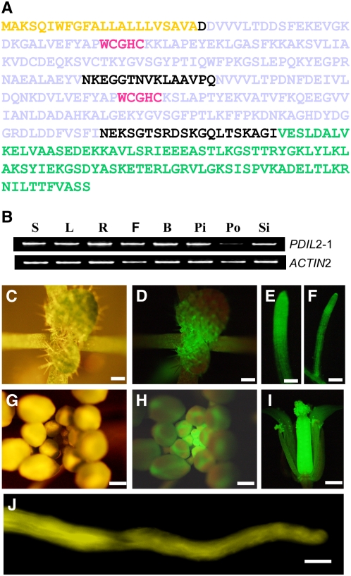
(A) Protein sequence of PDIL2-1. Yellow denotes the signal peptide (SP), gray denotes the two thioredoxin domains (TRX), red denotes the active sites, and green denotes the D domain.
(B) Expression of PDIL2-1 in different organs was examined by RT-PCR using gene-specific and control (ACT2) primers. S, seedlings; L, leaves; R, roots; F, opened flowers; B, vlosed flower buds; Pi, unfertilized pistils; Po, pollen; Si, siliques.
(C) to (J) Expression pattern determined by YFP fusion reporter (ProPDIL2-1:SP-eYFP-PDIL2-1).
(C) and (G) Vegetative and reproductive apical region.
(D) and (H) The same region under UV light (GFP filter).
(E), (F), and (I) Expression of PDIL2-1 in lateral and main roots and in a flower, respectively.
(J) In vitro–germinated pollen tube.
Bars = 500 μm in (C), (D), and (G) to (I), 100 μm in (E) and (F), and 10 μm in (J).

