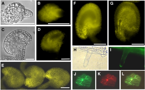
Figure 7.
PDIL2-1 Expression in Ovules and Subcellular Localization.
(A) and (B) Ovule of early stage 12 flowers under differential interference contrast (DIC) (A) or YFP channel (B). YFP signal is visible in integument cells and nucellus cells.
(C) and (D) Ovule of late stage 12 flowers under DIC (C) or YFP channel (D). YFP is visible in integument cells.
(E) Mature ovules of stage 13 flowers. YFP signal is visible in integument cells and is highly accumulated at the micropyle.
(F) An ovule of stage 13 flowers. Focus is on the plane of the embryo sac to show no signal inside of the embryo sac.
(G) A fertilized ovule with no YFP signal in the developing embryo.
(H) Root hair under transmitted light.
(I) ProPDIL2-1:SP-YFP-PDIL2-1 expression in root hairs.
(J) GFP-KDEL, an ER marker.
(K) PDIL2-1-tdTomato red fusion protein.
(L) Overlay of (J) and (K).
Bars = 20 μm in (A) to (D) and 50 μm in (E) to (G).

