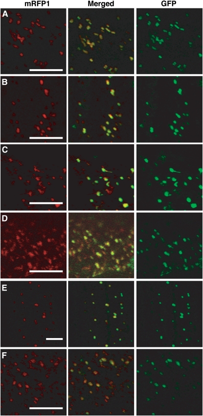
Figure 3.
Gm PNC1, At PNC1, and At PNC2 Are Targeted to Peroxisomes.
The polypeptides encoding Gm PNC1, At PNC1, and At PNC2 were fused at the N terminus or C terminus of mRFP1 under the control of the cauliflower mosaic virus 35S promoter. GFP-PTS1 was used as a marker of peroxisomes. Onion epidermal cells were used for the transient expression of combinations of GFP-PTS1 with mRFP1-Gm PNC1 (A), Gm PNC1-mRFP1 (B), mRFP1-At PNC1 (C), At PNC1-mRFP1 (D), mRFP1-At PNC2 (E), or At PNC2-mRFP1 (F). Confocal laser microscopy observation of the transiently expressed fluorescent proteins (RFP, red; GFP, green) was performed in epidermal cells. Center panels showed merged images. Bars = 10 μm.

