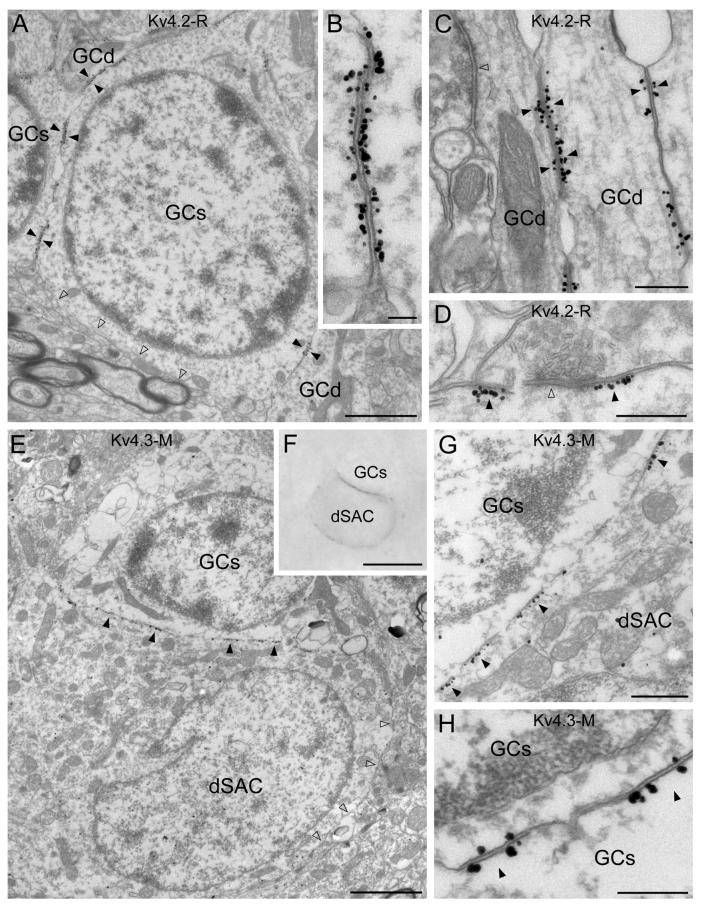
Fig. 3.
Electron microscopic immunogold localization reveals the subcellular distribution of the Kv4.2 and Kv4.3 subunits in the granule cell layer. (A) Immunogold particles for Kv4.2 subunit are clustered in membrane specializations (filled arrowheads) between granule cell somata (GCs) and dendrites (GCd). A part of the granule cell plasma membrane, which is not in direct contact with other granule cell processes (open arrowheads), contains a low density of labeling. (B) In such K+ channel-rich specializations, both apposing granule cell membranes are strongly labeled for the Kv4.2 subunit. (C) Kv4.2 subunit immunopositive junctions (filled arrowheads) are frequently observed between granule cell dendrites (GCd) in the GCL. Open arrowhead points to a synaptic junction. (D) Clustered immunolabeling for Kv4.2 subunit (filled arrowheads) is occasionally found around symmetrical synapses (e.g. open arrowhead) on a granule cell dendrite. (E and F) Clusters of gold particles for the Kv4.3 subunit (filled arrowheads) are observed in a deep short-axon cell (dSAC) contacting a granule cell soma (GCs). The high density of labeling is absent from other parts of the plasma membrane (open arrowheads in E). (F) Light microscopic image of the cells shown in E taken before EM sectioning. (G) A higher magnification image of the Kv4.3 subunit-rich specializations (filled arrowheads) between a dSAC and a granule cell soma (GCs). (H) Rare Kv4.3 subunit immunopositive junctions (filled arrowheads) between granule cell somata (GCs) are shown. Note that both apposing parts of the membrane specializations are labeled. Scale bars: 1.5 μm (A); 0.1 μm (B); 0.3 μm (C-E, G, H); 2 μm (E); 10 μm (F).

