Fig. 4.

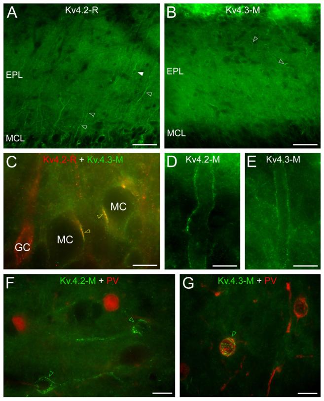
Distribution of the Kv4.2 and Kv4.3 subunits in the mitral cell and external plexiform layer. (A) Strongly Kv4.2 subunit immunolabeled dendrites (open arrowheads) dominate the homogeneous labeling of the neuropil of the external plexiform layer. A few immunopositive cell bodies are also observed (e.g. filled arrowhead) in the EPL. (B) A few Kv4.3 subunit immunopositive cell bodies (e.g. open arrowheads) stand out from the homogeneous labeling of the neuropil. (C) Double immunofluorescent labeling for the Kv4.2 and Kv4.3 subunits demonstrate their colocalization in clusters on mitral cells somata (yellow open arrowheads). (D and E) Distal parts of mitral cell apical dendrites are outlined by Kv4.2 (D) and Kv4.3 (E) subunit labeling. (F) Parvalbumin (red) immunonegative cell bodies are decorated by Kv4.2 subunit immunopositive clusters (green open arrowheads). (G) A parvalbumin immunopositive cell (green open arrowhead) of the EPL is decorated by strong Kv4.3 subunit positive clusters. Scale bars: 50 μm (A and B); 10 μm (C-G).
