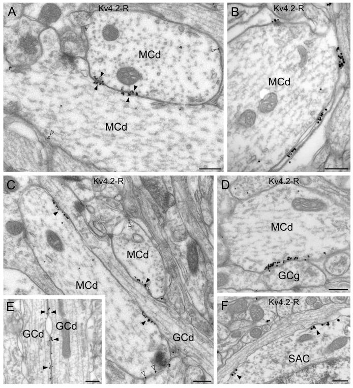
Fig. 5.
Electron microscopic immunogold localization of the Kv4.2 subunit in the mitral cell and external plexiform layers. (A) Clusters of immunogold particles for the Kv4.2 subunits are observed in membrane specializations (filled arrowheads) between two mitral cell dendrites (MCd). Note that gold particles are present in the membrane of both cells. A low intensity labeling was also found in MC lateral dendrites (open arrowheads). (B) Immunogold labeling is present in the glial sheet wrapping mitral cell apical dendrites (MCd). (C and D) A granule cell dendrite (GCd in C) and a gemmule (GCg in D) form strongly immunopositive membrane specializations with mitral cell dendrites (MCd). At these junctions, gold particles (filled arrowheads) are only found in the plasma membrane of the mitral cells. A lower density of labeling is also observed in these cells outside the specializations (open arrowheads). (E) Kv4.2 subunit immunopositive specializations are shown between granule cell dendrites (GCd) ascending into the EPL (filled arrowheads). (F) Kv4.2 immunopositive specializations (filled arrowheads) are observed in the soma of a short axon cell (SAC). Scale bars: 0.3 μm.

