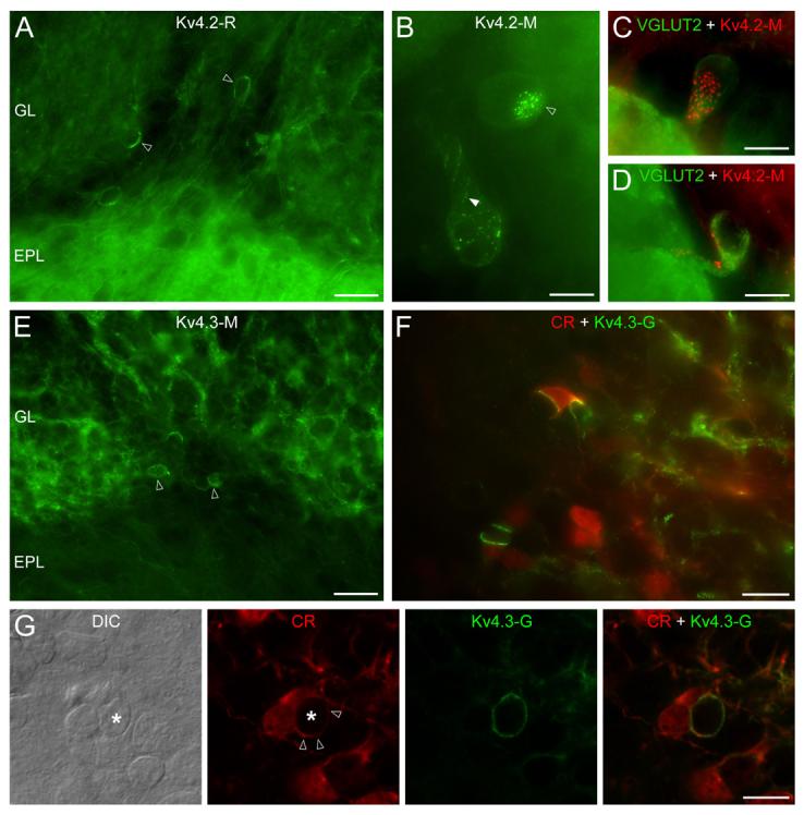
Fig. 6.

Distribution of the Kv4.2 and Kv4.3 subunits in the glomerular layer. (A) In addition to the weak neuropil labeling of the glomeruli, some juxtaglomerular cells (open arrowheads) are strongly Kv4.2 subunit immunopositive. (B) The uneven distribution of Kv4.2 subunit immunolabeling is revealed at higher magnifications. Small clusters are arranged either in disk-like (open arrowhead) or string-like (filled arrowhead) shapes. (C and D) Kv4.2 (red) subunit immunopositive cells are VGLUT2 (green) expressing external tufted cells. (E) Reticular-like labeling of the glomerular neuropil is found for the Kv4.3 subunit, in addition to a small number of immunopositive periglomerular cells (open arrowheads). (F) Double Kv4.3 subunit (green) and calretinin (CR: red) immunolabeling reveals that the intense Kv4.3 subunit clusters are often present on CR+ cells. (G) A strongly Kv4.3 subunit immunopositive process of a CR+ cell (open arrowheads) envelops another juxtaglomerular cell (*, note the nucleus on the DIC image). epifluorescent images (A and C-F); ‘extended focal image’ projection of 18 optical sections taken at 0.38 μm (B); single confocal sections (G); Scale bars: 25 μm (A and E); 20 μm (F); 10 μm (B, C, D and G).
