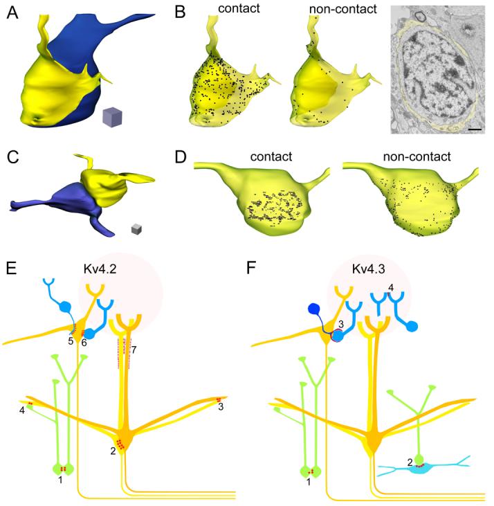
Fig. 8.

Three-dimensional reconstruction of Kv4.2 and Kv4.3 subunit immunopositive cells and schematic representation of the main cellular elements, containing K+ channel-rich junctions. (A) 3D reconstruction of a periglomerular cell soma (blue) enwrapped by a cap-like process (yellow). (B) Distribution of the Kv4.3 subunit in the plasma membrane of the process (transparent yellow). The density of the immunogold particles on the inner membrane of the cap, contacting the cell body (contact), is much higher than that on the outer membrane (non-contact). Gold particles labeling the Kv4.3 subunit are represented as black dots. An electron micrograph from the series used for the reconstruction is shown. Scale bar: 1 μm. (C) 3D reconstruction of two granule cell somata contacting each other. (D) Distribution of the Kv4.2 subunit in the plasma membrane of one of the granule cells (yellow cell). The labeling is more intense where the cell is in contact (contact) with the other granule cell (blue cell) compared to the rest of the somatic plasma membrane (non-contact). The length of each edge of the cubes: 1 μm. (E) Schematic representation of the clustered subcellular distribution of the Kv4.2 subunit in the MOB. Clusters of the subunit were found on both sides of junctions between: somata of granule cells (green, 1), mitral cells (yellow, 2) and dendrites of mitral cells (3). Asymmetrical enrichment of the Kv4.2 subunit was observed in: mitral cells contacting granule cell dendrites (4), external tufted cells (yellow) contacting periglomerular cell (blue) dendrites (5) and somata (6). The glial sheath surrounding the distal part of the mitral cell apical dendrites also showed clustered immunolabeling (7). (F) Schematic representation of the clustered subcellular distribution of the Kv4.3 subunit. Punctuate labeling of the subunit was found in both sides of conjunctions between the somata of a small number of granule cells (1). Clusters of the subunit were observed in: a subpopulation of deep short-axon cells (light blue) contacting granule cell somata (2), processes of a gliaform juxtaglomerular cell forming perisomatic caps around periglomerular cells (3), and in periglomerular cell dendrites contacting other dendrites in the glomeruli (4).
