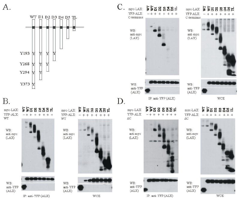
Figure 5.

The association of ALX with LAX maps to two distinct site within the LAX cytoplasmic tail. (A) C-terminal truncation mutants of LAX are shown, with the four tyrosines in the cytoplasmic tail of LAX that are sites of TCR-induces tyrosine phosphorylation denoted. (B-D) Expression plasmids for myc-tagged wt LAX or the LAX truncations shown in fig. 5A were co-transfected into 293T cells with YFP-tagged ALX constructs. WT ALX (B), ALX C (C), or ALX ΔC (D) proteins were used. Each experiment was otherwise performed as in Fig. 3. Note that the LAX truncations were expressed at a higher level than WT LAX as shown in the whole cell extract controls, and any increase in association with ALX appears proportional to the increased expression.
