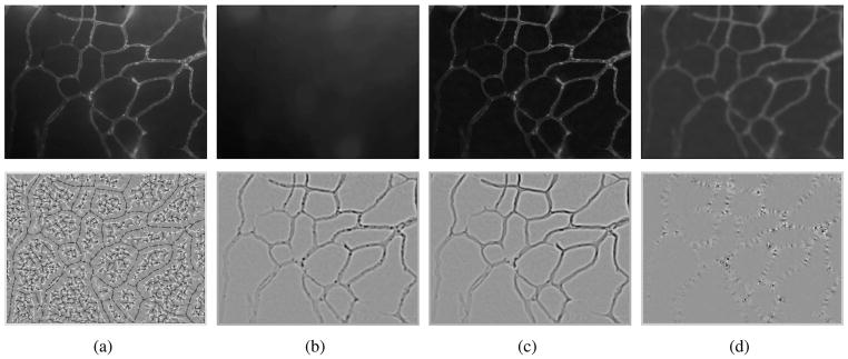
Fig. 1.
Row 1:Vessel enhancement(a) Original intensity map, (b) background estimation (opening), (c) background adjusted image (top-hat filter), (d) smoothed image (anisotropic diffusion filter + sqrt transformation). Row 2: Ridgeness measures(a) Mean curvature H, (b) Laplacian, (c) λ1, (d) λ2 (|λ1| ≥ |λ2|). Images in second row have been obtained by shifting the actual values. Dark: negative values, bright: positive values.

