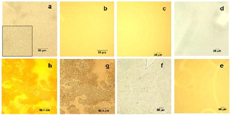
Figure 2.
Optical microscopy images of #5-Ce-meth-15sec with transmitted (a) and reflected (b) light, #5-Ce-meth-2min (c, reflected light), #5-Fe-meth-1min with transmitted (d) and reflected (e) light, #5-Fe-meth-5min (f, transmitted light), and #5-Fe-meth-30min with transmitted (g) and reflected (h) light. Inset in (a) shows a higher magnification image. The scale bar is 50 μm.

