
Fig. 2.
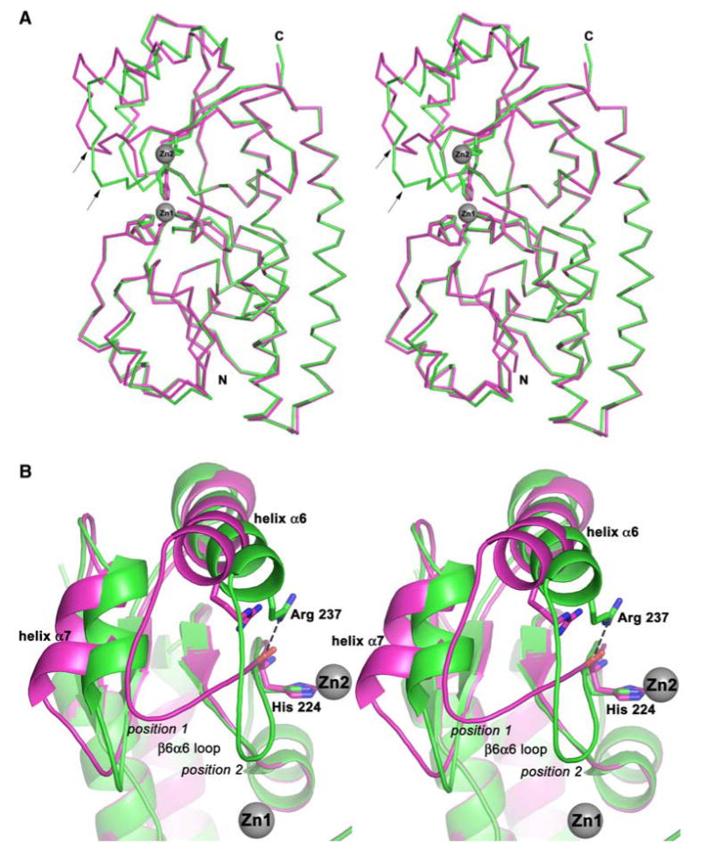
Structural comparison between ZnuA protein from Escherichia coli (Eco-ZnuA) with both metal binding sites occupied (ZnZnuA, molecule A, green) and Eco-ZnuA with one metal binding site occupied (CoZnuA, molecule E, magenta). a Stereo superposition of the Cα atoms. The β6α6 loop is labeled with arrows. b Closeup view of the conformational changes in the C-terminal domain. Residues His224 and Arg237 are shown as stick representations, and the Zn2+ ions are shown as gray spheres. The position of the Co2+ site is identical to that of Zn1