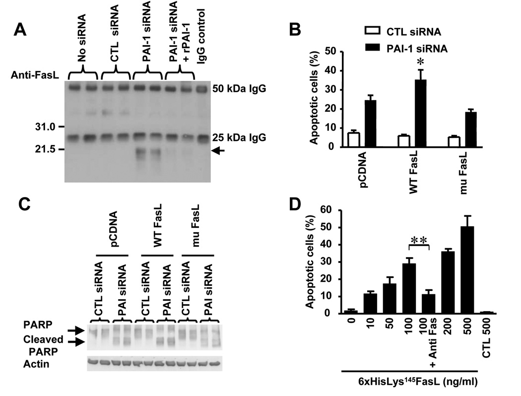
Figure 7. The 21.5 kDa Plasmin-generated FasL is Pro-apoptotic.
(A) The presence of the 21.5 kDa sFasL plasmin-generated fragment was detected by immunoprecipitation and Western blot analysis with an anti-FasL antibody in the CM of HBMEC treated as indicated on top.
(B) HBMEC were transfected with a pcDNA plasmid containing either a WT FasL or a mu FasL. Stable transfected cells were selected and re-transfected with a PAI-1 siRNA or a control siRNA and tested for apoptosis by FACS analysis after 72 hr. The data represent the means (± SD) of triplicate samples (* p< 0.05).
(C) Cells treated as described in (B) were examined for the presence of cleaved PARP by Western blot analysis.
(D) The recombinant 6xHisLys145FasL protein that corresponds to the 21.5 kDa plasmin-generated FasL obtained as shown in supplemental data Figure 2C was added at indicated concentrations to HBMEC for 48 hr and the percentage of apoptotic cells was measured by flow cytometry. An anti-Fas antibody (ZB4) was added at 500 ng/ml. The data represent the means (±SD) from triplicate samples (** p<0.005).

