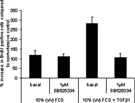
Figure 3.
PASMCs derived from iPAH patients were plated at equal cell densities in 96-well plates. Cells were starved for 48 hours before treatment with 0.625 ng/ml of TGF-β1 in growth media containing 10% (v/v) fetal calf serum. One μmol/L of SB525334 was added 15 minutes before the addition of TGF-β1. Proliferation was measured by BrdU incorporation after 6 days. The percentage of cells that were BrdU-positive was calculated and normalized to the average BrdU incorporation in untreated normotensive cells.

