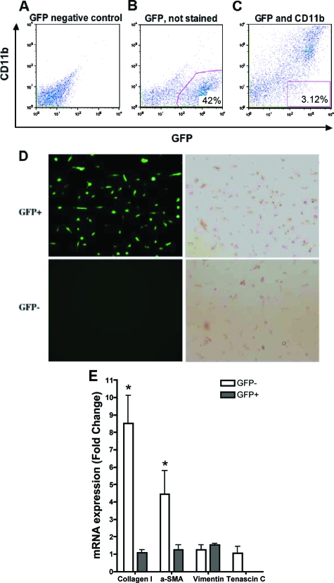
Figure 4.
Phenotypic analysis of purified GFP-positive lung fibroblasts. A: Wild-type (GFP-negative) control lung fibroblasts were sorted and used for gating purposes. B: GFP-positive cells were distinguished based on the calibration developed in A. C: Using the calibration developed in A and B, sorting of CD11b-negative lung fibroblasts. D: Microscopic analysis (fluorescence, left; phase contrast, right) revealed morphology consistent with fibroblasts in both the GFP-positive (top) and GFP-negative (bottom) cell populations. E: RNA from the purified GFP-positive and -negative cells was analyzed by real-time PCR for the following mRNA species: collagen I, α-smooth muscle actin, vimentin, and tenascin-C. Except for vimentin, which was equally expressed in both cell populations, expression of these genes was lower in the GFP-positive cells relative to the GFP-negative cells. Data are expressed as 2−ΔΔCT, using GAPDH as the reference and the GFP-negative cell signal as calibrator. N = 12 animals per group. *P < 0.05.

