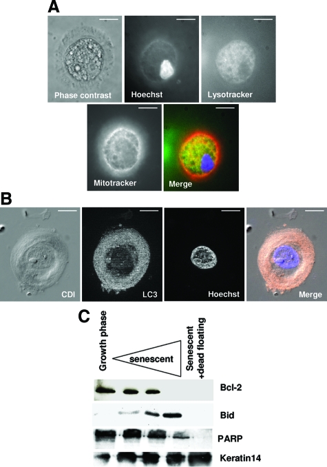
Figure 12.
Corpses degrade their mitochondria and nuclei in a central area full of autophagic vacuoles. A: Triple staining of a corpse with Lysotracker (green), Hoechst (blue) and Mitotracker (red). The three stains colocalize within a central area that occupies most of the corpse volume. The nucleus appears pycnotic. Scale bars = 10 μm. B: Corpse immunostained with an antibody against LC3 (pink), co-stained with Hoechst (blue), and analyzed by circular dichroism and with the ApoTome system. The four images represent one optical section. The central area is full of LC3-positive punctate structures and contains a damaged nucleus. Scale bars = 10 μm. C: Western blots. Cell extracts were performed with NHEKs in the growth phase and at different time points in the senescence plateau. Last lane: extracts from still-adherent cells plus dead floating cells.

