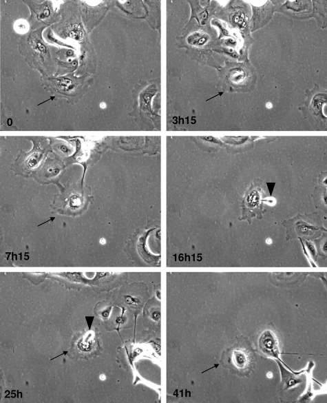
Figure 4.
and supplemental video, see http://ajp.amjpathol.org. Time-lapse phase contrast videomicroscopy of senescent cells undergoing death NHEKs at the senescence plateau were followed by videomicroscopy. Pictures were taken at 15-minute intervals for 24 to 48 hours. Some images taken at the indicated times were extracted for the figure. The senescent cell that will evolve into a corpse is indicated by an arrow. Note that the nucleus, at first, seems surrounded by a big structure (conspicuous at 3 hours 15 minutes). Then the dying cell detaches from the other cells but remains attached to the support for several hours. The dying cell expulses some dense material (arrowheads) several times in the course of the process.

