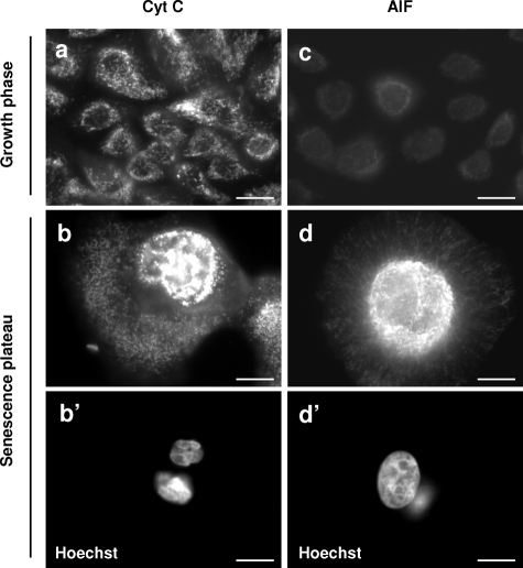
Figure 8.
The mitochondria of senescent keratinocytes are altered but do not release cytochrome C or AIF. NHEKs in the growth phase or at the senescence plateau were processed for immunofluorescence with specific anti-cytochrome C and anti-AIF antibodies. Both signals increase with senescence. The cytochrome C-positive structures change from radiating stick-shaped structures in growth-phase cells (a) to vesicular ones agglutinated in the vicinity of the nucleus in senescent cells (b and b′, note that the shown senescent cell contains two pycnotic nuclei). The AIF staining is very faint in young cells (c). It increases greatly in senescent cells; AIF-positive structures are very numerous and agglutinated around the nucleus (d and d′, note that the nucleus of the shown senescent cell is very large). Senescent keratinocytes do not show clear translocation of AIF into the nucleus or cytochrome C release into the cytosol (b and d). Scale bars = 10 μm.

