Figure 7.
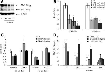
BimEL (23kD) and BimL (16kD) proteins are decreased after addition of asbestos to C10 lung epithelial cells (A, B) and up-regulated after silencing of PKD (C) or inhibition of ERK1/2 using U0126 (D). A: Representative Western blot and (B) quantitation of data. C: Quantitative data showing that BimEL (23kD) is increased in siPKD transfected cells. D: Quantitative data showing that U0126, but not SP600125, prevents asbestos-associated decreases in BimEL. The ERK1/2 inhibitor, U0126 (10 μmol/L), and JNK1/2 inhibitor, SP600125 (SP; 10 μmol/L) were added before asbestos as described in Materials and Methods. *P ≤ 0.05 in comparison with unexposed cells (0); †P ≤ 0.05 in comparison with siControl at same time point. Bars = Mean ± SEM.

