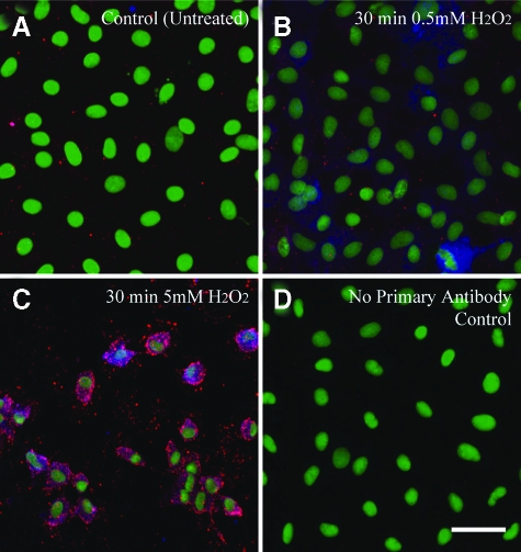
Figure 9.
Immunofluorescence experiments showing simultaneous induction of H2O2-induced pJNK1/2 and pERK1/2 signaling in C10 lung epithelial cells. (A) shows untreated confluent C10 cells. B and C show C10 cells exposed to 0.5 mmol/L and 5 mmol/L H2O2 for 30 minutes. (D) illustrates the lack of p-JNK1/2 or p-ERK1/2 staining in cells stained with secondary antibody alone. Nuclei are stained with Sytox Green. Red dots indicate p-ERK1/2 phosphorylation. All micrographs at original magnification ×400. Bar = 50 mmol/L.

