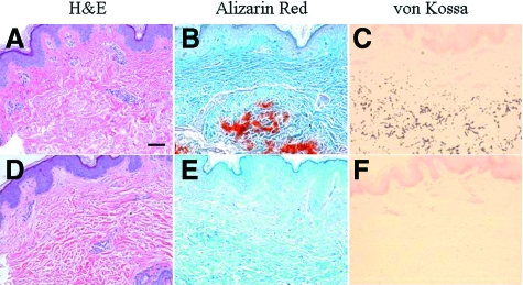
Figure 3.
Histopathology of the cutaneous lesions in the proband (A–C), in comparison with an unrelated healthy control (D–F). Staining with H&E (A,D) demonstrates basophilic abnormal elastic structures in the mid-dermis, and special stains (Alizarin Red and von Kossa) revealed that these elastotic structures are mineralized. Scale bar = 0.1 mm.

