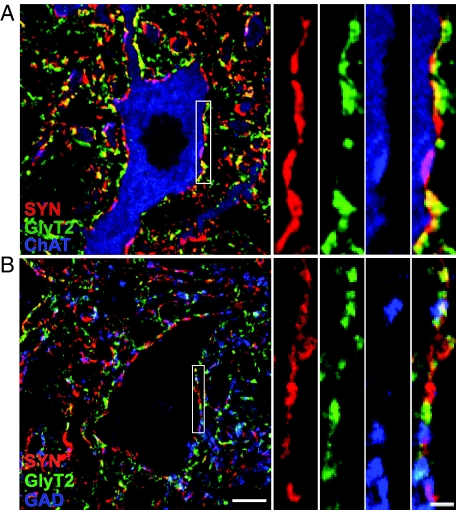
Figure 3.
Optical sections of lumbar motoneurons illustrating GlyT2- (green), GAD- (B; blue), and ChAT- (A; blue) positive boutons colocalized with synaptophysin (SYN; red) immunoreactive presynaptic terminals. A: Colocalization (yellow) of GlyT2 and synaptophysin, and colocalization (pink) of ChAT and synaptophysin around the soma and proximal dendrites of ChAT-positive motoneurons. B: Colocalization (yellow) of GlyT2 and synaptophysin, and colocalization (pink) of GAD and synaptophysin around the putative motoneurons. Three-way colocalization (white) of GlyT2-, GAD-, and synaptophysin immunoreactivity was also observed. Scale bar = 10 μm. The area delineated by white rectangular is shown of higher magnification at right. Scale bar = 2 μm.

