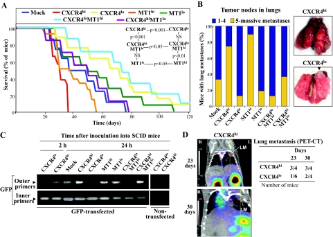
Figure 3.
CXCR4 and MT1-MMP are mutually required for in vivo melanoma metastasis. A: Survival curves of mice (n = 10) inoculated into the tail vein with the indicated transfectants, and statistics from the different curves (N.S., nonsignificant). B: Degree of lung colonization by melanoma transfectants (left), and representative lung metastases from CXCR4hi (34 days, massive metastasis) and CXCR4lo (70 days) mice. Arrow indicates a metastatic tumor node. C: Melanoma transfectants were transiently transfected with pEGFP-C1 vector, and subsequently intravenously inoculated into SCID mice. After the indicated times, GFP expression in lungs was determined by nested PCR. Negative controls with nontransfected cells are also shown. D, left: SCID mice were inoculated with CXCR4hi or CXCR4lo melanoma transfectants and subjected to PET-CT analyses. Co-registered PET and CT studies were superimposed. Coronal sections from a CXCR4hi mouse at days 23 and 30 are shown. Intersections between lines correspond to the center of lung metastases (LM) (R, right; L: left). D, right: Data indicate number of mice with lung metastases, mostly one to two tumor nodes.

