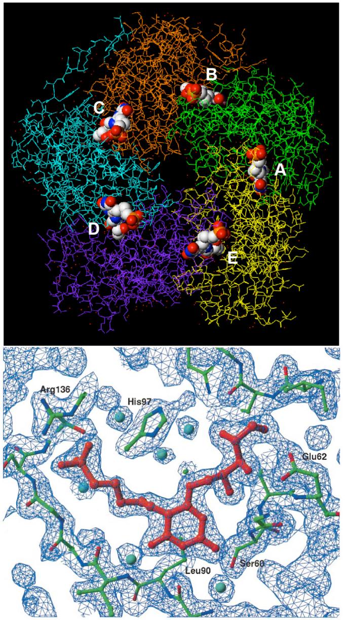
Figure 7.
Top: crystal structure (PDB ID: 1EJB) of S. cerevisiae lumazine synthase pentamer with bound ligand 15. The five subunits are shown in stick form, while the five ligand molecules are shown as space-filling models color coded according to atom type. The N-termini in the subunits exist in different conformations in the crystalline state. The five binding sites, which contain amino acid residues from adjacent monomers, are labeled A-E. Bottom: stick model and electron density map of inhibitor 15 bound in the active site (22).

