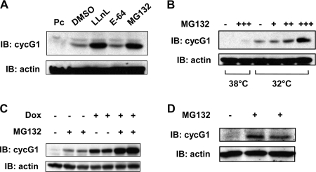
FIG. 2.
Exogenously and endogenously expressed cyclin G1 protein can be stabilized by inhibitors of the 26S proteasome. (A) NIH 3T3 cells were transfected with constructs expressing murine cyclin G1 or with empty pcDNA3 vector (Pc) for 20 h, after which cells were left untreated or treated with LLnL (25 μM), E-64 (50 μM), or MG132 (50 μM) for 5 h. Equivalent amounts of cell lysates (30 μg) were separated by 10% SDS-PAGE and immunoblotted with cyclin G1 (178) or actin antibodies, as indicated. DMSO, dimethyl sulfoxide. (B) Clone 6 rat cells expressing temperature-sensitive mutant p53 (A135V) were maintained at 38°C. Cells were shifted to 32°C for 5 h and then either left untreated or treated with 5 μM (+), 10 μM (++), or 50 μM (+++) MG132 for an additional 5 h. Cells were lysed and separated by 10% SDS-PAGE followed by immunoblotting with the indicated antibodies, as in panel A. Parallel cultures maintained at 38°C were left untreated or treated with 50 μM MG132 for 5 h and then treated as described above. (C) HeLa Tet-on cells (clone 2), engineered to express tetracycline/doxycycline-inducible human cyclin G1 as described in Materials and Methods, were plated for 24 h, followed by treatment with or without doxycycline (2 ng/ml) for 20 h. Cells were then either left untreated or treated with 25 μM MG132 for an additional 5 h prior to lysis. Equivalent amounts of cell extracts (30 μg) were separated by SDS-PAGE and immunoblotted with anti-human cyclin G1 polyclonal antibody H-46 or anti-actin antibody. (D) H1299 cells were left untreated or treated with 50 μM MG132 for 4 h prior to lysis and separation of cell extracts (50 μg) by 10% SDS-PAGE followed by immunoblotting with H-46 and antiactin antibodies.

