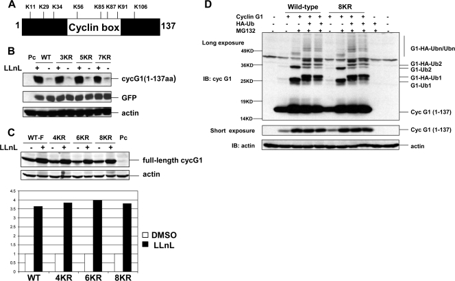
FIG. 5.
All lysines in the N terminus of cyclin G1 are dispensable for its degradation and ubiquitination. (A) Positions of the eight lysine residues in the N-terminal 137 amino acids of the murine cyclin G1 protein. (B) NIH 3T3 cells were transfected with constructs expressing an N-terminal version (residues 1 to 137) of wild-type or lysine substitution (3KR, 5KR, and 7KR) mutant cyclin G1 or with empty pcDNA3 vector (Pc) for 28 h and then were left untreated (−) or treated (+) with LLnL (+) (25 μM) for 12 h. Cell extracts (30 μg) were separated by 12% SDS-PAGE and immunoblotted with anti-cyclin G1 (178), anti-GFP, or antiactin antibodies. For N-terminal (residues 1 to 137) cyclin G1 substitution mutants, 3KR represents K11R, K29R, and K34R mutations; 5KR represents K11R, K29R, K34R, K56R, and K85R mutations; and 7KR represents K11R, K29R, K34R, K56R, K85R, K87R, and K91R mutations. (C) NIH 3T3 cells were transfected with constructs expressing full-length versions of wild-type or N-terminal lysine substitution (4KR, 6KR, or 8KR) mutant cyclin G1 or with empty pcDNA3 vector (Pc) for 28 h and then were left untreated (−) or treated with LLnL (+) (25 uM) for 12 h. Cell extracts (30 μg) were separated by 10% SDS-PAGE and immunoblotted with anti-cyclin G1 (178) or antiactin antibodies. The graph shows the increase (fold change) in wild-type and mutant cyclin G1 proteins compared with untreated levels, as analyzed by an Odyssey scanning system. For full-length cyclin G1 substitution mutants, 4KR represents K11R, K29R, K34R, and K56R mutations; 6KR represents K11R, K29R, K34R, K56R, K85R, and K87R mutations; and 8KR represents K11R, K29R, K34R, K56R, K85R, K87R, K91R, and K106R mutations. (D) NIH 3T3 cells were transfected with wild-type or mutant (8KR) N-terminal (residues 1 to 137) cyclin G1 as in panel C, with or without HA-ubiquitin, for 24 h, followed by treatment with MG132 (25 μM) for 5 h. Cell lysates were separated by 12% SDS-PAGE and immunoblotted with anti-cyclin G1 (178) or antiactin antibodies. The middle panel shows a shorter exposure of the cyclin G1 portion of the blot. G1-Ub1, G1-Ub2, G1-HA-Ub, and G1-HA-Ub2 are described in the legend to Fig. 3B.

