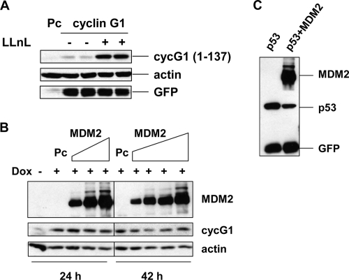
FIG. 7.
Cyclin G1 degradation by the 26S proteasome is p53 and MDM2 independent. (A) MDM2−/− p53−/− (2KO) cells were transfected with the N-terminal cyclin G11-137 construct or empty pcDNA3 vector (Pc) for 28 h and either left untreated or treated with LLnL (25 μM) for 12 h. Cell extracts (30 μg) were separated by 12% SDS-PAGE and analyzed by immunoblotting. (B) HeLa Tet-on cells expressing human cyclin G1 (clone 2) were left untreated or treated with doxycycline (2 ng/ml) and, at the same time, were transfected with increasing amounts of Flag-HDM2 for 24 h (0.5 μg, 1.0 μg, and 2.0 μg) or 42 h (0.2 μg, 0.5 μg, 1.0 μg, and 2.0 μg) or with empty pcDNA3 vector (Pc). Cell lysates were separated by 10% SDS-PAGE and analyzed by immunoblotting with anti-Flag (M2), anti-human cyclin G1 (H-46), or antiactin antibodies. (C) HeLa Tet-on cells (clone 2) without doxycycline were transfected with 0.3 μg of p53-expressing plasmid, with or without 1.5 μg of HDM2-expressing plasmid, for 24 h. Cell lysates were separated by 10% SDS-PAGE and analyzed by immunoblotting with anti-Flag (M2), anti-p53 (DO-1), or anti-GFP antibodies.

