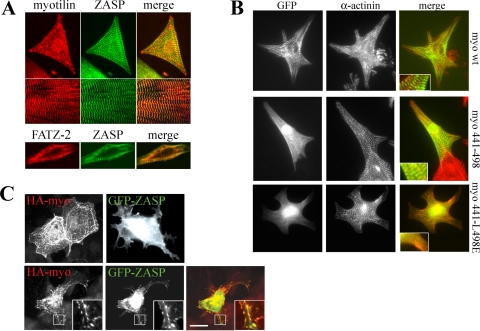
FIG. 2.
Myotilin and ZASP/Cypher colocalize in muscle cells, and myotilin is involved in targeting ZASP/Cypher. (A) Rat neonatal cardiomyocytes (upper panel) and sections of skeletal muscle (middle panel) were stained myotilin (left, red) and ZASP/Cypher (middle, green). The bottom panel shows staining of cardiomyocytes for FATZ-2 (left, red) and ZASP/Cypher (middle, green). On the right, merged images are shown. (B) GFP-myotilin, GFP-myotilin 441-498, and GFP-myotilin 441-L498E were transfected to rat neonatal cardiomyocytes and stained for α-actinin as a Z-disc marker. Wild-type and C-terminal myotilin is targeted to the Z-discs in the sarcomere, whereas the C terminus with L498E substitution is not. The insets show larger magnifications of the merged images. (C) HA-myotilin and GFP-ZASP were transfected to COS-7 cells either (upper panel) or together (lower panel). Myotilin was detected with anti-HA antibody and Alexa-594-coupled secondary antibody (red). Single transfected myotilin induces thick filaments, whereas ZASP/Cypher is uniformly distributed in the cytoplasm. In double-transfected cells, ZASP/Cypher is targeted to the filaments, where it colocalizes with myotilin. wt, wild type. Scale bar, 5 μm.

