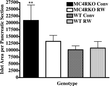
Figure 6.
At termination of the experiment, pancreatic tissue was immediately excised from mice after death. The tissue was fixed in formalin and imbedded in paraffin for sectioning. Nonserial sections of each sample were then subjected to hematoxylin and eosin staining and insulin immunostaining. Anti-insulin-stained islets were analyzed for β-cell area, number, and endocrine to exocrine tissue ratios using Zeiss Axiophot image analysis software. Individual islet cell areas of 3 nonserial sections of each sample were analyzed. Area values for 3 to 4 animals/group were averaged and compared. Bars represent averages ± se; n = 3–4 mice/group. ▪, MC4R KO sedentary group; □, MC4R KO RW group; ▩, WT littermate sedentary group; ▥, WT RW group. **P < 0.01; two-way ANOVA with Bonferroni post hoc tests.

